Are you seeking a method to reveal the intricacies of cellular processes? The calcium flux assay might just be the breakthrough you’ve been searching for. By measuring the movement of calcium ions within cells, this assay unravels the essential dynamics of cellular signalling. Using fluorescent dyes, scientists can visualise and quantitate the changes in calcium ion levels with great precision. This technique not only deepens understanding of cellular functions but also plays a critical role in drug discovery and disease research. Explore how the calcium flux assay is transforming our approach to understanding cellular behaviour in this comprehensive guide.
Understanding Calcium Flux Assay Basics
A calcium flux assay is a pivotal laboratory technique for measuring the movement of calcium ions within cells. This process is essential for understanding a wide array of cellular processes. The assay employs fluorescent dyes that bind selectively to calcium ions, enabling researchers to visualise and quantify changes in intracellular calcium levels. By observing these changes, scientists gain insights into various cellular functions and signalling pathways, underscoring the assay’s importance in biomedical research.
- Drug Discovery: Evaluates the effects of potential therapeutics on calcium signalling pathways.
- Neurotransmitter Release: Assesses how neurons communicate by monitoring calcium ion dynamics.
- Muscle Contraction: Studies the role of calcium in muscle fibres during contraction and relaxation.
- Immune Response: Investigates calcium-dependent processes in immune cell activation and response.
- Hormone Secretion: Analyses the calcium role in the release of hormones from endocrine cells.
Calcium flux assays are integral to advancing our understanding of cellular mechanisms. Their ability to track calcium ion movement offers valuable insights into cellular responses and interactions. This makes them indispensable in both basic research and applied biomedical studies, facilitating breakthroughs in understanding and treating various physiological and pathological conditions.
Methodology of Calcium Flux Assay
The calcium flux assay is a meticulous laboratory technique designed to measure intracellular calcium levels. The procedure relies on fluorescent indicators that selectively bind to calcium ions. These indicators allow researchers to visualise and quantify calcium dynamics in real-time. The use of fluorescence microscopes or plate readers is crucial to detect and measure the emitted fluorescence. Ensuring proper experimental controls throughout the process is essential for obtaining accurate and reliable results.
Preparation and Loading of Calcium Indicators
Selecting the appropriate fluorescent dye is critical for the success of the assay. The chosen dye must have high specificity and affinity for calcium ions to provide accurate measurements. Preparation involves dissolving the dye in a suitable solvent, often dimethyl sulfoxide (DMSO), and diluting it to the required concentration. Samples, typically consisting of cells in a culture medium, are then incubated with the dye. This incubation period allows the dye to permeate the cell membranes and bind to intracellular calcium. Proper washing steps are necessary to remove unbound dye and reduce background fluorescence, thereby enhancing the signal’s clarity.
Measurement Techniques
Fluorescence detection is primarily conducted using either fluorescence microscopes or plate readers. Fluorescence microscopes offer the advantage of visualising calcium dynamics in individual cells, providing detailed spatial information. Plate readers, on the other hand, are ideal for high-throughput analysis, enabling the simultaneous measurement of multiple samples. Both methods involve exciting the calcium-bound fluorescent indicators with a specific wavelength of light, causing them to emit fluorescence. The intensity of this emission is directly proportional to the concentration of intracellular calcium, allowing quantitative analysis. Calibration with known calcium concentrations is essential to ensure data accuracy and reliability.
| Step | Description |
|---|---|
| Preparation and Loading | Selecting appropriate fluorescent dyes, preparing samples, and ensuring efficient dye loading and washing. |
| Measurement Techniques | Utilising microscopy or plate readers to detect fluorescence and quantify calcium levels. |
Equipment and Materials Required for Calcium Flux Assay

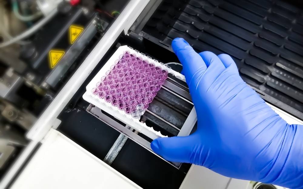
Conducting a calcium flux assay requires precise equipment and materials to ensure accurate measurement and analysis. Essential equipment includes fluorescence plate readers or flow cytometers. These tools are pivotal for detecting and quantifying the fluorescence emitted by calcium-bound dyes, allowing researchers to assess intracellular calcium levels effectively. Fluorescence microscopy is also frequently employed to visualise calcium dynamics within cells, providing detailed spatial information.
- Calcium-Sensitive Dyes: Used to bind calcium ions and emit fluorescence for detection.
- Fluorescence Plate Reader: Measures fluorescence intensity across multiple samples efficiently.
- Flow Cytometer: Allows detailed analysis of individual cells’ calcium levels.
- Microscope Slides and Coverslips: Needed for mounting samples for microscopy.
- Dimethyl Sulfoxide (DMSO): Solvent used to dissolve calcium-sensitive dyes.
- Culture Medium: Provides the necessary environment for maintaining cell viability during the assay.
Proper calibration and preparation of these materials are indispensable for the assay’s success. Calibration ensures that the fluorescence measurements accurately reflect the calcium concentrations, while meticulous reagent preparation, including dye concentration and solvent use, minimises variability and enhances data reliability. These steps are crucial to obtaining consistent and meaningful results from calcium flux assays, facilitating insights into cellular mechanisms.
Applications of Calcium Flux Assays in Biomedical Research
Calcium flux assays hold a pivotal role in pharmacological research, particularly in drug discovery. These assays provide critical insights into how potential therapeutics affect calcium signalling pathways within cells. By measuring the movement of calcium ions, researchers can determine the efficacy and mechanism of action of new compounds, facilitating the development of drugs that precisely target cellular responses. This capability is invaluable in assessing drug safety and efficacy at an early stage, which significantly accelerates the drug development process.
Drug Discovery and Screening
How do calcium flux assays contribute to drug discovery? They enable researchers to evaluate how new drugs interact with calcium channels and receptors. This interaction is crucial because calcium ions serve as secondary messengers in many signal transduction pathways. By observing changes in calcium ion concentrations, researchers can infer how a drug modulates these pathways. This information helps in identifying compounds that can effectively alter cellular behaviour, making calcium flux assays a cornerstone in the pharmacological evaluation of new therapeutic agents.
Cellular Function Studies
Calcium flux assays are also extensively used to study various cellular functions. For instance, in muscle contraction, calcium ions play a vital role in the sliding filament mechanism that enables muscle fibres to contract. By using calcium flux assays, researchers can explore how different stimuli affect muscle contraction at the cellular level. Additionally, these assays are instrumental in understanding immune responses, as calcium signalling is crucial for the activation and function of immune cells. By measuring calcium flux, scientists can gain insights into how immune cells respond to pathogens or other stimuli, which is essential for developing therapies to modulate immune responses.
The versatility of calcium flux assays in biomedical research cannot be overstated. By providing a quantitative measure of cellular responses to various stimuli, these assays help unravel complex biological processes. This ability to elucidate cellular mechanisms makes them indispensable tools in advancing our understanding of health and disease, ultimately contributing to the development of novel therapeutic strategies.
Challenges and Considerations in Conducting Calcium Flux Assays
Conducting calcium flux assays involves several challenges, particularly in terms of assay sensitivity and specificity. One of the primary concerns is selecting fluorescent dyes that effectively bind to calcium ions without cross-reacting with other ions, which can lead to inaccurate results. Maintaining cell viability throughout the assay is also critical, as compromised cells may not accurately reflect calcium dynamics. Minimising background fluorescence is essential to ensure the observed signals are truly representative of calcium activity rather than artefacts.
- Choice of Fluorescent Dyes: Select dyes with high specificity and minimal cross-reactivity.
- Optimisation of Dye Concentration: Ensure the dye concentration is sufficient for detection but not cytotoxic.
- Sample Preparation: Conduct thorough washing steps to reduce background fluorescence.
- Temperature Control: Maintain consistent temperature to prevent variations in cellular responses.
- Calibration Standards: Use calibration curves to correlate fluorescence intensity with calcium concentrations accurately.
To enhance assay performance and accuracy, it is vital to adopt a meticulous approach. This includes validating the specificity and sensitivity of fluorescent dyes in preliminary assays and ensuring optimal dye loading by refining incubation times and concentrations. Employing robust calibration techniques is also crucial to translate fluorescence readings into meaningful calcium ion concentrations. By addressing these considerations, researchers can obtain reliable and reproducible data, crucial for advancing understanding in cellular studies.
Interpretation and Analysis of Calcium Flux Assay Results
The interpretation and analysis of calcium flux assay results hinge on precise fluorescence intensity measurements. These measurements are critical because they provide a quantitative assessment of intracellular calcium ion concentrations. How is this achieved? By employing fluorescence indicators that emit light when bound to calcium ions, the fluorescence intensity directly correlates with the concentration of calcium present within the cells. Accurate measurement of this fluorescence is essential to determine the impact of various stimuli on cellular calcium dynamics.
- Data Collection: Gather raw fluorescence intensity data from the assay, ensuring measurements are taken at consistent intervals.
- Baseline Correction: Subtract background fluorescence to obtain an accurate measure of calcium-dependent fluorescence.
- Calibration: Use known calcium concentrations to create a standard curve, allowing translation of fluorescence intensity into calcium concentrations.
- Kinetic Analysis: Evaluate changes in fluorescence over time to understand the dynamics of calcium ion flux in response to stimuli.
Understanding the biological context is crucial for interpreting calcium flux assay results accurately. This involves recognising the specific cellular processes under investigation and how calcium ions function within those processes. For instance, in muscle cells, calcium plays a pivotal role in contraction; thus, changes in fluorescence intensity can indicate alterations in muscle function. Similarly, in neurons, calcium flux is integral to neurotransmitter release. By thoroughly understanding these biological systems, researchers can draw meaningful conclusions from the data, enhancing insights into cellular responses and interactions.
Final Words
Exploring calcium flux assays highlights their crucial role in deciphering cellular processes. By measuring calcium ion movement through fluorescence, researchers can gain valuable insights into cell function.
The methodology and equipment are detailed, ensuring accurate and reliable results in various biomedical applications, such as drug discovery and cellular function studies.
Challenges like dye specificity and cell viability are manageable with careful planning, enhancing the assay’s reliability.
Interpreting results requires understanding biological contexts, promising advancements in research and therapeutic developments.
Harnessing the power of calcium flux assays promises significant contributions to scientific understanding and innovation.

James is a music journalist with a passion for live performances, festival culture, and emerging artists. He covers everything from stage setups to behind-the-scenes stories of major music events.